
Why in News? For those trying to look inside the human body without surgery, magnetic resonance imaging (MRI) is an indispensable tool.
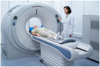
What is MRI? MRI is a non-invasive diagnostic procedure that is used to obtain images of soft tissues (that hasn’t become harder through calcification) within the body. It is widely used to image the brain, the cardiovascular system, the spinal cord and joints, various muscles, the liver, arteries, etc. Its use is particularly important in the observation and treatment of certain cancers (like prostate) and to track neurological conditions including Alzheimer’s, dementia, epilepsy, and stroke.
When was it Developed? In the early 1970s, techniques for MRI were developed. Later, it was refined by Lauterbur and Mansfield. Their work led to the Nobel Prize in Medicine in 2003, highlighting MRI's pivotal role in diagnostics.
How does MRI Work? MRI machines use the magnetic properties of hydrogen atoms in the body to create images. The machine includes a superconducting magnet creating a stable magnetic field, aligning hydrogen atom spins. A radiofrequency pulse is emitted, exciting excess atoms. When the pulse stops, these atoms emit energy, detected and converted into signals by a receiver. These signals are processed by a computer to generate detailed 2D or 3D images of the scanned body part.
What are the Pros of MRI? An MRI scan can practically image the body from all useful directions. Once the magnetic fields are taken away, the atoms in the scanned part don’t remain affected. This means, MRI scans do not pose any threats to the body.
What are the Cons of MRI? MRI machines are expensive and diagnostic facilities pass this cost on to its patients- each scan often costs Rs 10,000 or more. As MRI technique use strong magnetic fields, individuals with metallic implants (including pacemakers) may not be able to undergo MRI scans. Also, a scan’s effects on pregnant women aren’t well-studied.
