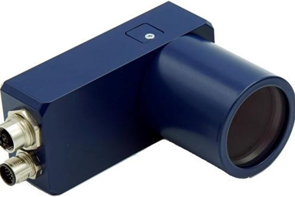
About Neuromorphic camera:
- A neuromorphic camera mimics the way the human retina converts light into electrical impulses.
- How does it work?
- In a typical camera, each pixel captures the intensity of light falling on it for the entire exposure time the camera focuses on the object. All these pixels are pooled together to reconstruct an image of the object.
- In neuromorphic cameras, each pixel operates independently and asynchronously, generating events or spikes only when there is a change in the intensity of light falling on that pixel.
- This generates sparse and lower amounts of data compared to traditional cameras, which capture every pixel value at a fixed rate, regardless of whether there is any change in the scene.
- This allows a neuromorphic camera to "sample" the environment with much higher temporal resolution because it is not limited by a frame rate like normal cameras and also performs background suppression.
- Neuromorphic cameras have a very high dynamic range ( >120 dB) which means they can be used in different conditions ranging from a very low-light environment to very high-light conditions.
What is meant by diffraction limit in Optical Microscopy?
- The resolution of a microscope is proportional to the size of its objective and inversely proportional to the wavelength of light being observed.
- In 1873 the German physicist Ernst Abbe discovered how microscopes were limited by the diffraction of light. He revealed that the resolution of a microscope is not controlled by the instrument’s quality but by the wavelength of light used and the aperture of its optics.
- Due to this phenomenon, a microscope cannot resolve two objects located closer than λ/2NA, where λ is the wavelength of light and NA is the numerical aperture of the imaging lens. This is known as the diffraction limit.
- Thus diffraction limits the ability of the microscope to distinguish between two objects divided by a lateral distance of less than half the wavelength of light used to image the sample.