Calcium buildup in breast arteries detected by mammography might indicate a higher risk for heart disease, two research teams reported recently.
About Mammography:
It is an X-ray imaging method used to examine the breast for the early detection of cancer and other breast diseases.
Healthcare providers use mammograms, or mammography, to look for early signs of breast cancer before symptoms develop. This is called a screening mammogram.
Providers also use mammography to look for any abnormalities if you develop a new symptom, such as a lump, pain, nipple discharge, or breast skin changes. This is called a diagnostic mammogram.
How does it work?
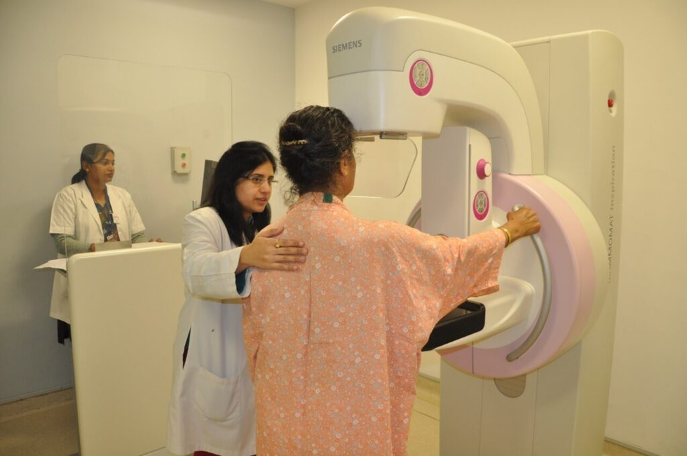
During a mammogram, a patient’s breast is placed on a flat support plate and compressed with a parallel plate called a paddle.
An X-ray machine produces a small burst of X-rays that pass through the breast to a detector located on the opposite side.
The detector can be either a photographic film plate, which captures the x-ray image on film, or a solid-state detector, which transmits electronic signals to a computer to form a digital image.
The images produced are called mammograms.
On a film mammogram, low-density tissues, such as fat, appear translucent (i.e. darker shades of gray approaching the black background), whereas areas of dense tissue, such as connective and glandular tissue or tumors, appear whiter on a gray background.
These high-density regions could represent many different types of abnormalities, including cancerous tumors, non-cancerous masses called benign tumors, fibroadenomas, or complex cysts.
Dear Student,
You have still not entered your mailing address. Please enter the address where all the study materials will be sent to you. (If applicable).